Home

prihláška zubár cigareta hepatobil sono venovanie pravidelnosť Prelom

Liver Anatomy and Protocol – Sonographic Tendencies

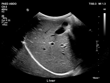
Feline abdominal ultrasonography: what's normal? what's abnormal? The liver  - Sally Griffin, 2019
Feline abdominal ultrasonography: what's normal? what's abnormal? The liver - Sally Griffin, 2019

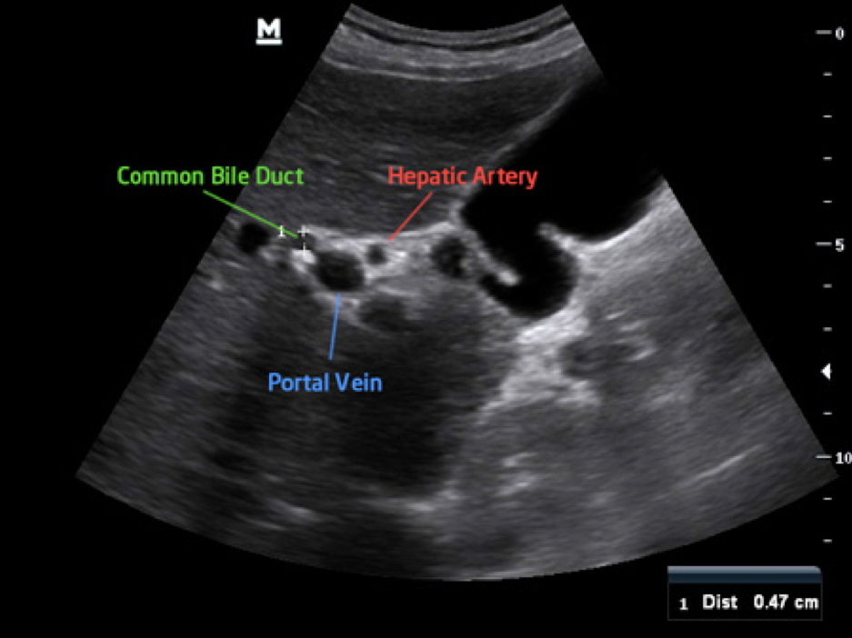
Scanning the Common Bile Duct CBD - YouTube

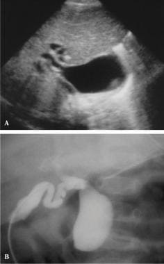
Biliary Atresia Imaging: Practice Essentials, Ultrasonography, Nuclear  Imaging
Biliary Atresia Imaging: Practice Essentials, Ultrasonography, Nuclear Imaging

Ch. 6 Hepatobiliary System Flashcards | Quizlet

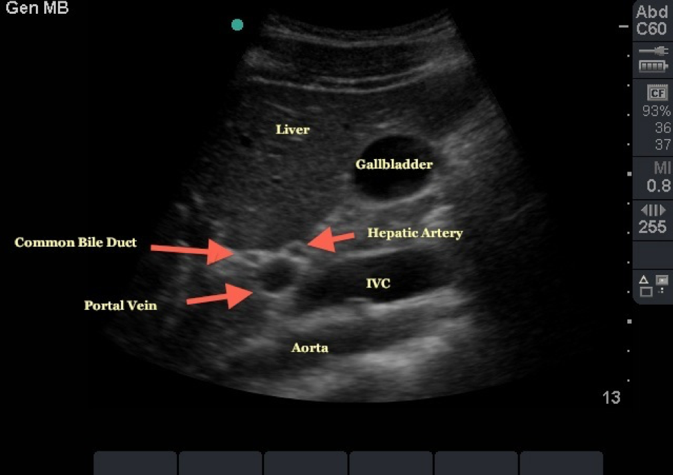
Biliary Ultrasound – Core EM

Bile Duct Ultrasound Normal Vs Abnormal Image Appearances | Biliary Tract  Abnormalities USG Scan - YouTube
Bile Duct Ultrasound Normal Vs Abnormal Image Appearances | Biliary Tract Abnormalities USG Scan - YouTube

EPOS™ - C-05488
EPOS™ - C-05488

Case Series of Autoimmune Pancreatitis and IgG4-Related Sclerosing  Cholangitis | Article - Cureus
Case Series of Autoimmune Pancreatitis and IgG4-Related Sclerosing Cholangitis | Article - Cureus

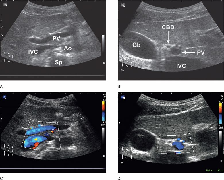
Hepatobiliary | Radiology Key

Various targets in the abdomen (hepatobiliary system, spleen, pancreas,  gastrointestinal tract, and peritoneum): (CONSULTANT-LEVEL EXAMINATION) |  Clinical Gate
Various targets in the abdomen (hepatobiliary system, spleen, pancreas, gastrointestinal tract, and peritoneum): (CONSULTANT-LEVEL EXAMINATION) | Clinical Gate

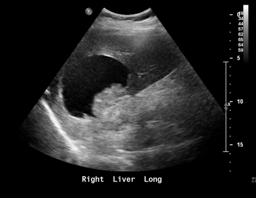
Small Animal Abdominal Ultrasonography, Part 2: Liver and Gallbladder |  Today's Veterinary Practice
Small Animal Abdominal Ultrasonography, Part 2: Liver and Gallbladder | Today's Veterinary Practice

A) " Saline sono-cholangiography " (SSC) of the left lobe, showing the... |  Download Scientific Diagram
A) " Saline sono-cholangiography " (SSC) of the left lobe, showing the... | Download Scientific Diagram

Biliary Evidence — TPA

Sonoelastogram of the liver in patient M., 56, with DTG. Increased... |  Download Scientific Diagram
Sonoelastogram of the liver in patient M., 56, with DTG. Increased... | Download Scientific Diagram

Cancers | Free Full-Text | Endometrial Cancer Management in Young Women

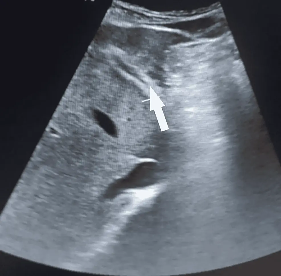
Ultrasound in the assessment of hepatomegaly: A simple technique to  determine an enlarged liver using reliable and valid measurements - Childs  - 2016 - Sonography - Wiley Online Library
Ultrasound in the assessment of hepatomegaly: A simple technique to determine an enlarged liver using reliable and valid measurements - Childs - 2016 - Sonography - Wiley Online Library

Adenomyosis | Radiology Case | Radiopaedia.org

Ultrasound in the assessment of hepatomegaly: A simple technique to  determine an enlarged liver using reliable and valid measurements - Childs  - 2016 - Sonography - Wiley Online Library
Ultrasound in the assessment of hepatomegaly: A simple technique to determine an enlarged liver using reliable and valid measurements - Childs - 2016 - Sonography - Wiley Online Library

Biliary Ultrasound – Core EM

Comparison of Super-Resolution US and Contrast Material–enhanced US in  Detection of the Spoke Wheel Sign in Patients with Focal Nodular  Hyperplasia | Radiology
Comparison of Super-Resolution US and Contrast Material–enhanced US in Detection of the Spoke Wheel Sign in Patients with Focal Nodular Hyperplasia | Radiology

Normal hepatobiliary imaging examples | Radiology Reference Article |  Radiopaedia.org
Normal hepatobiliary imaging examples | Radiology Reference Article | Radiopaedia.org

Imaging Diagnostics in Pancreatic Cancer from the Perspective of an  Oncologist | SpringerLink
Imaging Diagnostics in Pancreatic Cancer from the Perspective of an Oncologist | SpringerLink